Description
Confocal is just that simple
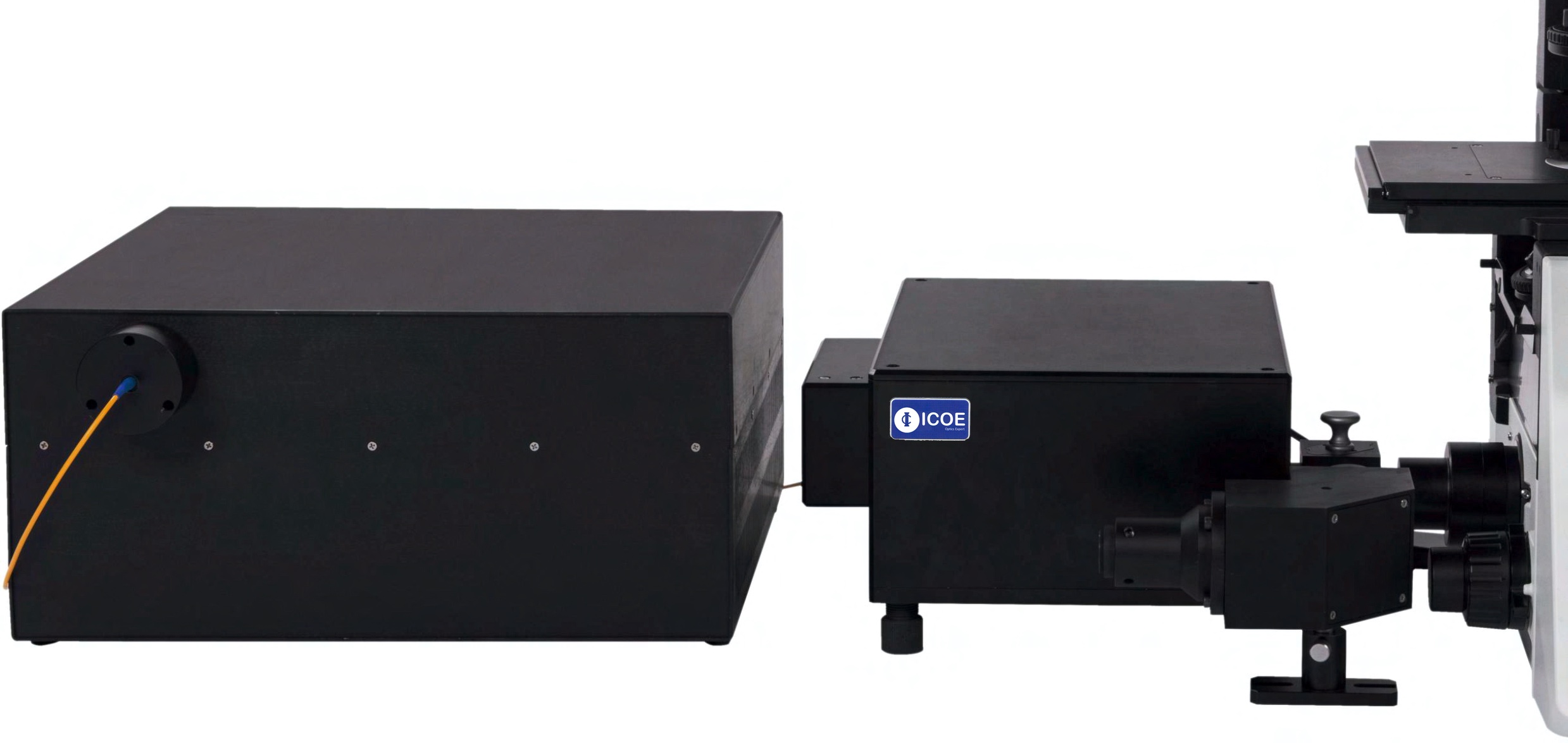
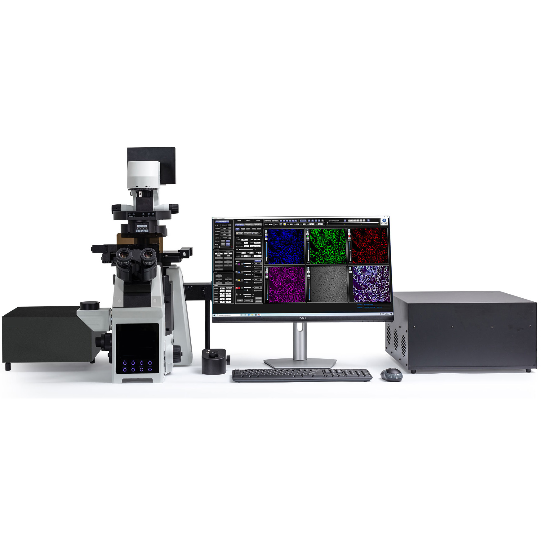
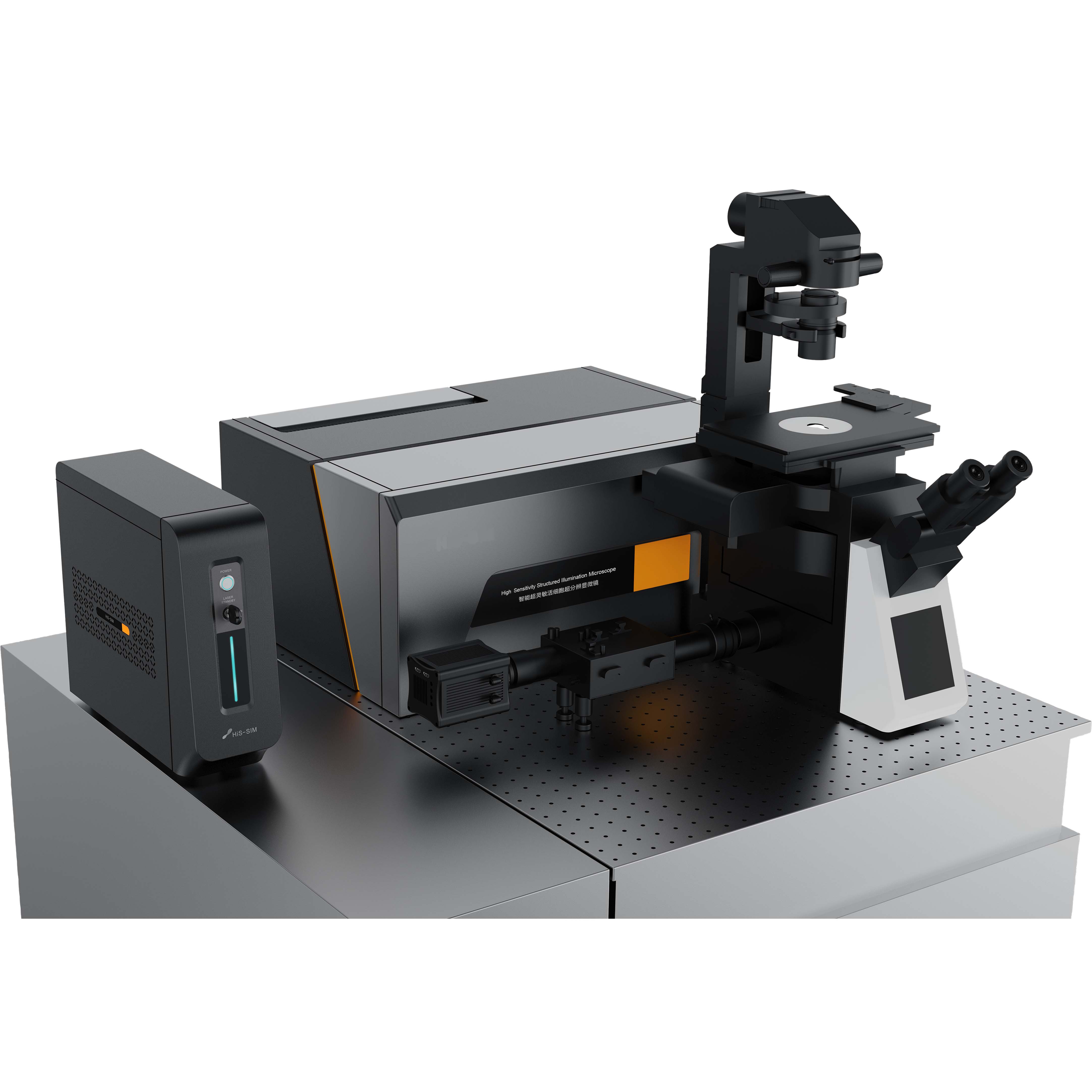
CSIM200 is a confocal imaging product developed with the design concept of simplicity and efficiency, which can help researchers easily obtain high quality confocal images.
The unique optical path structure design of CSIM200 puts the imaging pinhole behind, which not only ensures the collection efficiency of fluorescence signals, but also improves the filtering of non-focus plane signals, and improves the image signal-to-noise ratio and axial resolution. The self-developed signal amplification circuit meets the requirements of confocal imaging for high detection sensitivity and large dynamic range. Compared with previous confocal system images, the acquired confocal image background has lower noise, darker background and richer details.
Through the standard C interface, CSIM200 can be connected to any brand and model of research-grade microscopes, and the original wide-field fluorescence microscope can be quickly upgraded to a confocal imaging system. The operating system is simple, intuitive, and easy to use, and novices with zero confocal experience can easily obtain high-quality confocal images.
Simple, Efficient and Stable
The innovative optical path design of CSIM200 simplifies the optical path structure, reduces the influence of devices on system stability, and optimizes the shape and size of the confocal pinhole, which can block more non-focal plane signals than traditional confocal systems, and is closer to Ideal confocal.
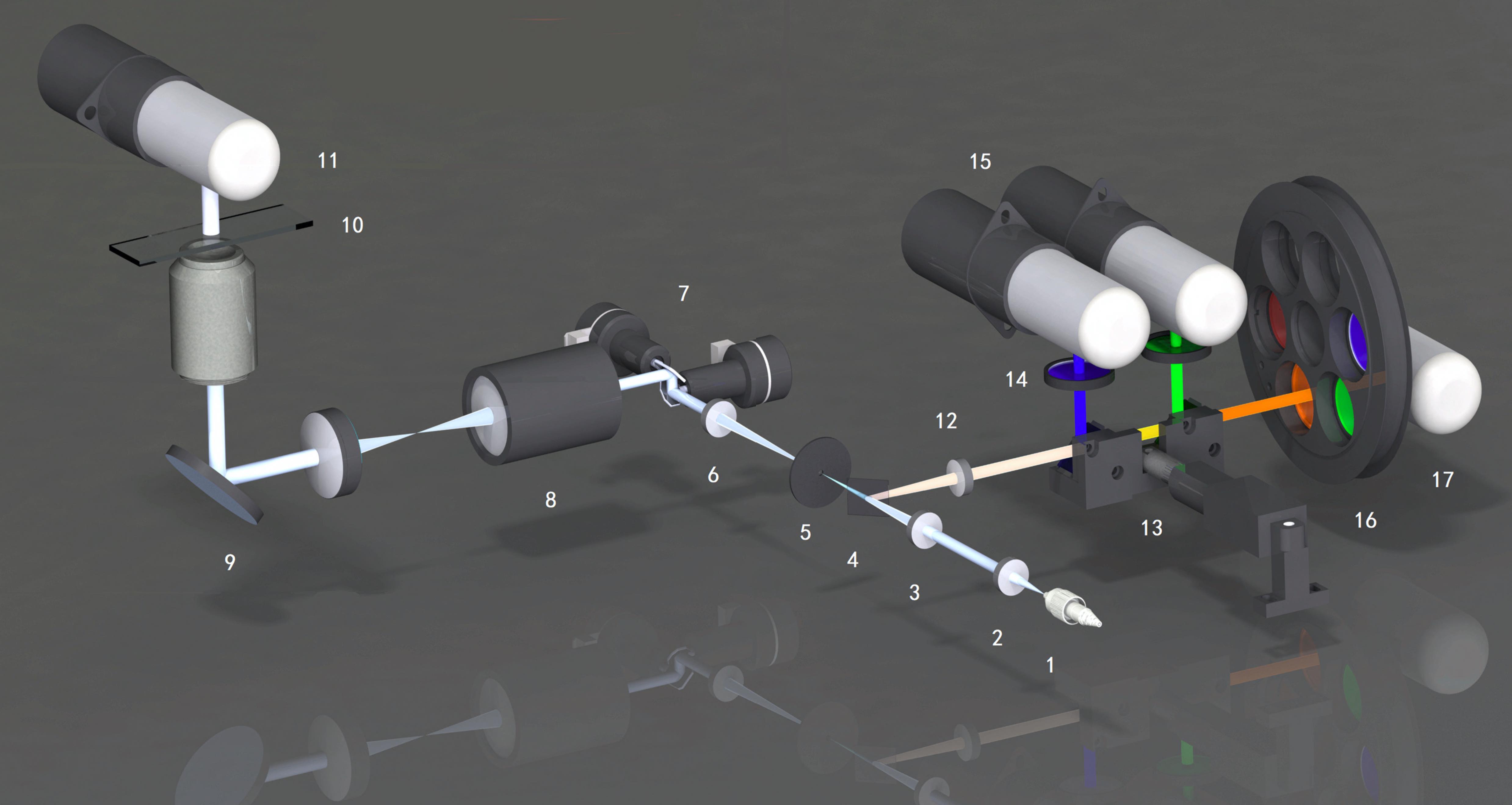
| 1. Optical fiber (laser) | 6. Pinhole adapted collimating lens | 11. DIC PMT | 16. Filter wheel |
| 2. Fiber optic collimator | 7. XY scanning galvanometer | 12. Detection collimator | 17. Fluorescent PMT |
| 3. Focusing lens | 8. Scanning lens | 13. Secondary dichroic beam splitter | |
| 4. Primary dichroic beamsplitter | 9. Microscope | 14. Emission filter | |
| 5. Pinhole | 10. Sample | 15. Fluorescence PMT |
Product Features
1. Confocal Pinhole Unit
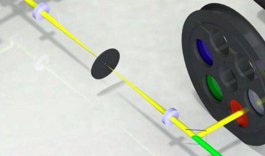
In order to reduce the slight displacement of various components in the optical path, the projection of the excitation point and the pinhole on the sample cannot be well overlapped (conjugated), and the pinhole blocks the fluorescence, resulting in a decrease in detection efficiency. Traditional confocals use long focal lengths. The lens magnifies the intermediate image and the pinhole, which is several times larger than the Airy disk on the imaging surface of the microscope. Although such a design ensures the detection efficiency of the fluorescence, it also reduces the efficiency of the pinhole blocking the non-focal plane signal from entering the detector, which reduces the Z-axis resolution of the final confocal image.
The CSIM200 makes the fluorescence emitted by the sample go back along the laser light path and pass through the pinhole, and then uses a dichroic mirror to separate the fluorescence from the laser light path and guide it to the detector, which minimizes the interference caused by component displacement and does not require the system The intermediate image and pinhole are magnified, and the intermediate image and pinhole match the Airy disk of the microscope at a ratio of 1:1. Signal-to-noise confocal images with better axial resolution than conventional confocals.
*For the upgrade plan of the variable pinhole, please consult us.
2. Detection Controller Unit
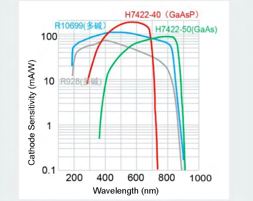
The detection unit of CSIM 200 is equipped with a 6-hole motorized filter wheel and a single high-sensitivity multi-alkali photomultiplier tube (MA PMT, QE≥25%@500nm) pre-installed with 4 high-performance interference filters , the system can quickly and automatically complete multi-color fluorescence co-imaging to obtain high-quality multi-color fluorescence confocal images. In addition, the standard differential interference (DIC) unit can obtain high-resolution differential interference images for fluorescence localization analysis.
3. Signal Acquisition Circuit
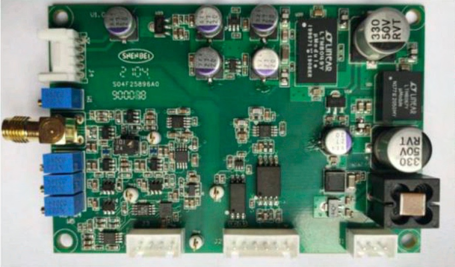
The signal amplification circuit of the CSIM200 detection unit is based on 20 years of analog circuit development experience of our company, and specially developed a new circuit for the characteristics of single-point scanning imaging. It uses fluorescence acquisition and electrical signal amplification different from traditional confocal systems. The method improves the acquisition efficiency of fluorescence signals by at least an order of magnitude, and also solves the problem of low dynamic range caused by the use of cross-group amplifiers in traditional confocal systems, so that the acquired images have greater contrast and richer detail.
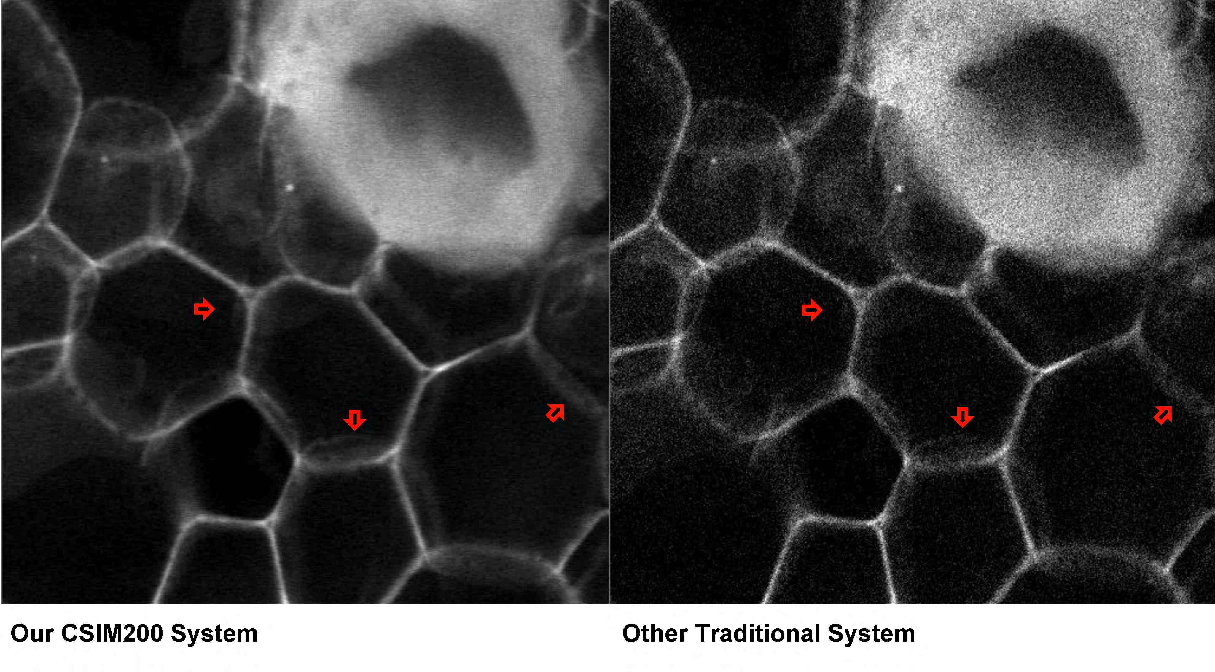
* For more filter options and upgrade solutions for GaAsP PMT, please contact us.
Software Function
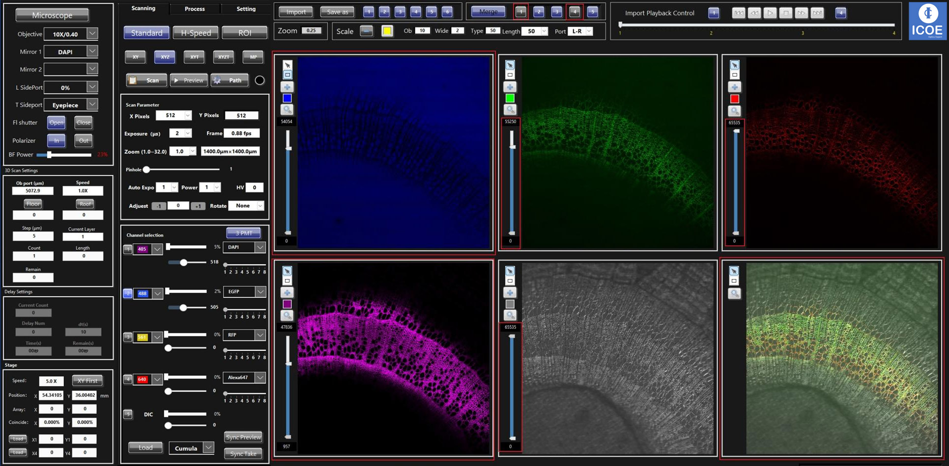
CSIM200 is equipped with imaging software with Chinese interface, which is easy to learn. Anyone can quickly master the imaging process and easily obtain multi-color two-dimensional confocal images.
The software can also automatically complete imaging experiments such as 3D imaging, time-lapse imaging, 3D time-lapse imaging and multi-point imaging, and can perform scale correction and addition, filter processing, Z-Stack and large image splicing and other processing and analysis, and supports output in multiple formats image, automatically save scan parameters.
Includes Differential Interference Interference (DIC) unit, motorized focus unit and motorized stage
CSIM200 Imaging Function
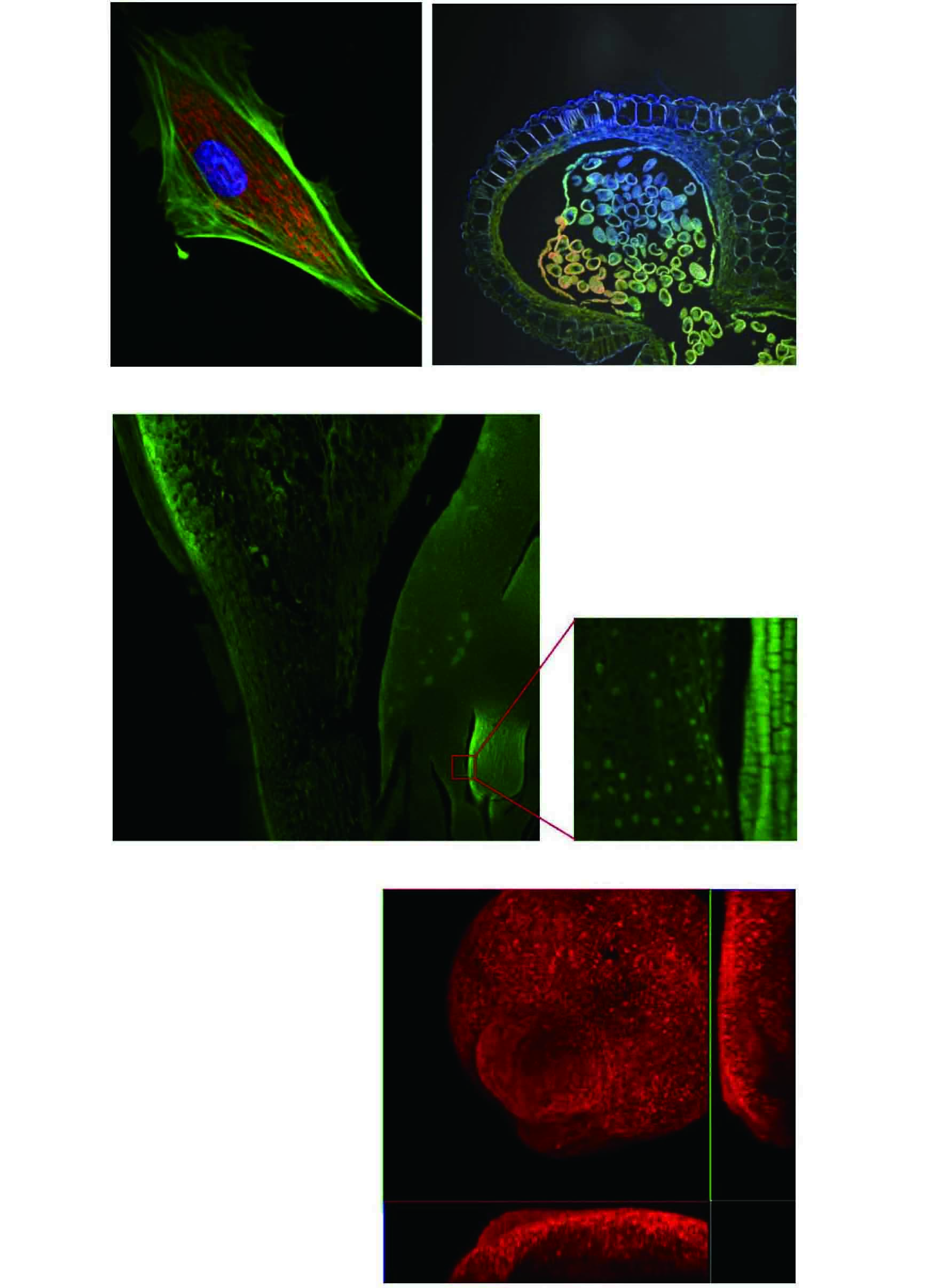
CSIM200 supports single-channel or multi-channel two-dimensional imaging (XY), three-dimensional imaging (XYZ), four-dimensional imaging (XYZT) and multi-site scanning.
* multi-channel imaging
* DIC
* Fluorescence Overlay
* Z-axis scanning 3D imaging (XYZ)
* 3D imaging (XYZT)
* Puzzle
CSIM200 System Application
Good confocal slice scanning effect, very friendly to thicker brain slice samples. It can be used to study the interaction of nerve cells and neural network research.
Model organisms
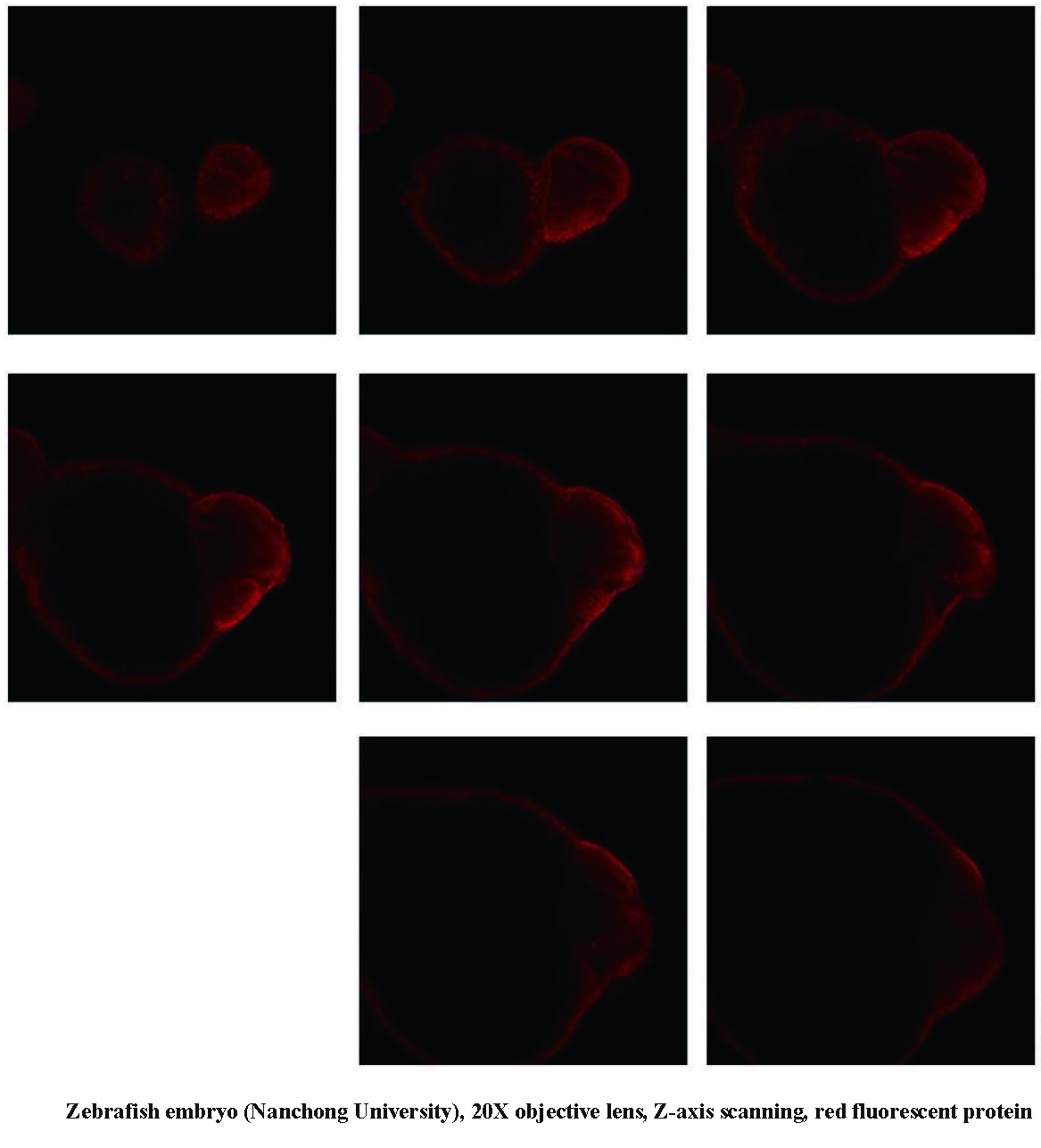
It can be used to observe model organisms such as zebrafish, Drosophila, nematodes and Arabidopsis, etc. Through large-field imaging, three-dimensional structural images of the entire sample are obtained through jigsaw puzzles, and the development process is recorded; through the observation of endogenously expressed fluorescent proteins, the dynamic changes in the fine structure of macromolecules are tracked.
Pharmacology and Pathology Research
In drug therapy research, obtain and quantify fluorescence data from single-label or multi-label fluorescent staining samples, screen and record the expression of fluorescent molecules, and obtain dynamic localization information and concentration information of labeled molecules. Morphological and qualitative fluorescence observations were performed on immunofluorescence-labeled pathological tissue sections.
Nervous System Research
Good confocal slice scanning effect, very friendly to thicker brain slice samples. It can be used to study the interaction of nerve cells and neural network research.
Subcellular Structure Research
Imaging live cells or fixed multi-fluorescently labeled samples to study fluorescence colocalization, dynamic properties or spatial relationships of two or more target proteins. For example, observe macromolecular structures such as mitochondria, endoplasmic reticulum, or cytoskeleton, and study the relationship between target molecules, organelles, and cytoskeleton during cell migration.
Tissue Slice Culture Research
Confocal can be very flexible and intuitive to observe the morphology through three-dimensional slice reconstruction. It is suitable for observing multicellular samples such as tissues, blood vessels, muscle fibers and artificial hearts in three-dimensional culture in vitro, tracking the rapid growth process of simulated muscle contraction, blood flow, whole organism or tissue culture, while retaining structural information.
Plant, Microbial Research
Confocal is also suitable for the observation and research of samples such as plant leaves, roots, bacteria, and prokaryotes. For example, related studies on plant photoperiod-related proteins, the motility of cyanobacterial cilia, and the tropism of bacteria.
Product Models & Specifications
Our CSIM200 is compatible with Major Brands’ Microscope Platform (Such as Olympus, Zeiss, Lecia, Nikon etc.)
As high compatibility of CSIM200’s Laser Confocal Imaging System, it can be installed with major brands’ microscope platform, such as Olympus, Zeiss, Lecia, Nikon etc, which provides a wealth of choices to users. All the purpose just for providing user best application solutions according to their preferences. If user already have one of those microscope platform in his laboratory, you can just buy one CSIM200, and it can be simply installed into the microscope platform to achieve confocal up-gradation, which can greatly help users when they have limited budgets.
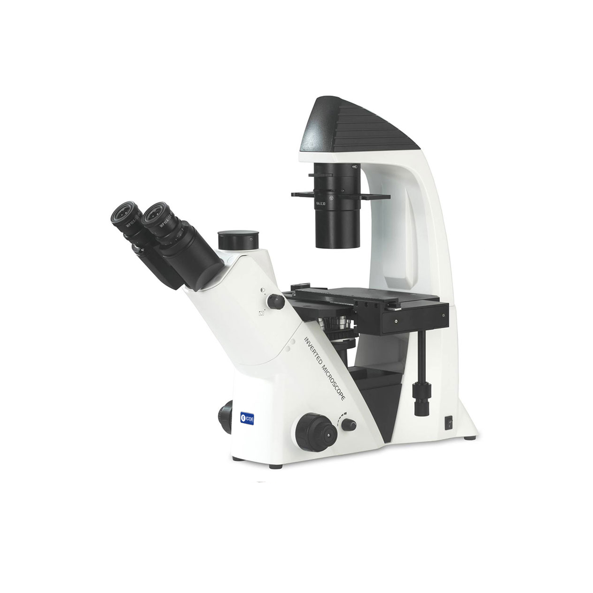
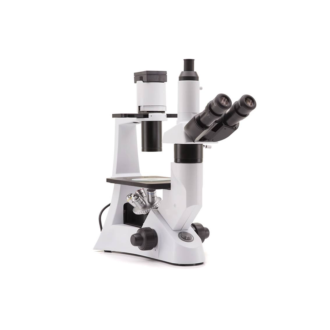
The Microscope Platform for ICF980A

